 Laboratory of Anatomy, Biomechanics and Organogenesis
In-vivo registration of both electrogoniometry and medical imaging: development and implementation
This research is improvements of the previous method to permit both scientific and clinical in-vivo applications. Main improvements were related medical imaging and landmark location. Low-dose computerized tomography was adopted to achieve reduced total dose absorption by the subject. In order to perform registration location of anatomical landmarks were used instead fiducial landmarks inserted into the bone structure. Accurate landmark definitions were written to reduce discrepancy between discrepancy between both virtual and manual palpation. Further development of the new method included on-line registration of the 3D bone models during joint kinematics data collection for visual feedback and quality checking of the collection procedure. Further motion data processing used both anatomical and helical data representation. The method was applied on the right ankle of one volunteer. Two trials were collected for reproducibility purposes. Results were satisfactory and in agreement with the literature.
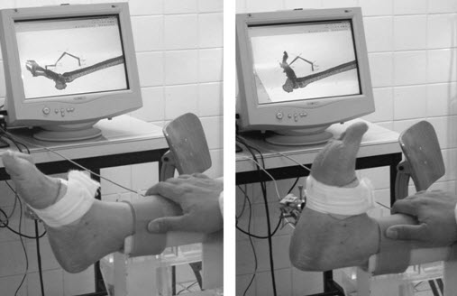
Two snapshots of the joint kinematics data collection. On-line registration of the subject's 3D bone models and 3D virtual goniometer model are displayed on the screen located in the background. Left image : ankle joint in plantarflexion. Right image : ankle joint in dorsiflexion. Observe the on-line registration on the screen. |
|
Need more information? Please contact ![]() !
!