Knee joint kinematics, thigh muscles moment arms and tibial proximal cancellous bone alteration after distal femoral osteotomies
Lower limb disorders have been considered as a factor inducing gonarthrosis and the three-dimensional effect of the surgical correction is not well reported yet. The long term clinical outcomes of this kind of surgery are sometimes not satisfactory. The aim of this research project to better understands the 3D effect of in-vitro distal femoral osteotomies applied following the three anatomical planes.
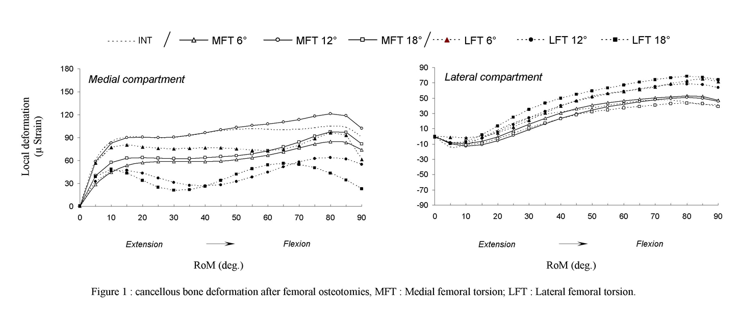
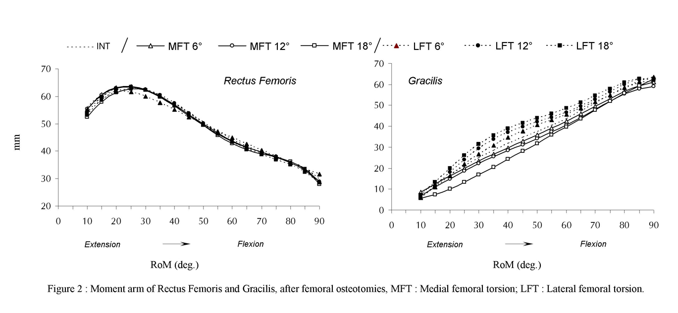
This research developed a method using the strain gauge to assess the proximal cancellous bone deformations. Knee joint kinematics was collected using a 3D electrogoniometer and the thigh muscles moment arms were estimated by the tendon excursion method using some Linear Variable Differential Transformer.
Prior to specimens’ preparation, each specimen has been fully CT-scanned to obtain 3D bone models. During specimen dissection, small spherical markers (diameter: 4mm) were inserted into the lower limb bones. These Spherical markers were digitalized on the 3D bone models (from the CT images) and the centroid of these spherical markers were digitalized using a Faro arm to get the 3D coordinates in the experimental setting.
This methodology allows us to record some biomechanical data in relation to the surgical procedure. Actually all these biomechanical data are implemented to ameliorate our computational anatomical data set.
In a close future we will be able to predict the alterations of the biomechanics of many anatomical tissues in relation to surgical procedure. The computational anatomy will help the surgeon in their decision making.
This methodology could be applied to any others joints.
Top image: cancellous bone deformation. Bottom image: thigh muscle moment arms.
|
|
Need more information? Please contact ![]() !
!
 Laboratory of Anatomy, Biomechanics and Organogenesis
